Posts

Showing posts from 2017
Image
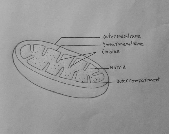
How to draw DNA The DNA has twisted ladder or double helical structure. According to Watson and Crick DNA molecule consists of two polynucleotide chains wrapped helically around each other, with the sugar- phosphate chain on the outside forming ribbon-like backbone of double helix. Purines and pyrimidines on the inside of the helix projecting between two sugar phosphate backbones as transverse bars. The two polynucleotide strands are held together by hydrogen bonds between specific pairs of Purines and Pyrimidines.  Now let's see how to draw DNA accurately.  1.Draw two vertical parallel lines as shown with pencil. 2. Draw a twisted wave touching two lines on either sides. 3. Draw another wavy curve as shown. Start the second curve  a bit near the first curve to get proper grooves of DNA. 4. Make ribbon shape on  the strands as shown. Make sure t
Image
How to draw chloroplast Chloroplast is cell organelle which is seen only in plant cells and algal cells. A typical chloroplast is in biconvex shape. It has outer membrane and inner membrane. The inner membrane encloses viscous fluid called stroma. In stroma there is network of  several piled thylakoids. Chloroplast is food synthesizing organelle. It prepares food called starch by process called  Photosynthesis .   Step 1: Draw a double convex shape as shown.   Step 2: Draw one more curve upon,  following the contours of below curve.   Step 3: Draw about three faint reference  lines inside as shown. These lines help in drawing grana thylakoids in proper orientation.   Step 4: Draw about three to five grana along the reference lines as shown. Step 5: Connect the grana thylakoids by grana lamellae as shown. Put several neat dots to represent stroma of chlorop
Image
How to draw Mitochondria Mitochondria  is cell organelle which generates power required for cell and hence called `Power house of the cell". They are sac like structures present in the cytoplasm of the cells. They may be of various shapes like thread, spherical or club. Mitochondria have two compartments an inner compartment and an outer compartment.  The viscous substance in the inner compartment is called Matrix. The matrix is surrounded by a membrane called Inner membrane. The inner membrane is thrown into several folds called Cristae. Now let`s draw mitochondria.   Step 1: Draw a rough sausage shape with free hand in faint curve. Do not stop the curve in the middle, continue until you meet the starting point.  Step 2: To give a sliced sausage look, draw one more curve as shown. Line inner side with another faint curve as shown.   Step 3: Erase the inner line leaving the faint impression and mark the positions of cristae wit
Image
How to draw a nerve cell ( Neuron) Nerve cell is functional unit of nervous system.It has mainly two parts the Cyton and Axon.Cyton have several dendrites emerging from surface. Cyton gives rise to Axon from a triangular structure called axon hillock. Axon of nerve cells sometimes extend to 1 mt length in nerves of leg. Cyton something looks like a star so, it can be drawn from a basic star shape.  Step 1: Draw two triangles superimposed as shown resembling a star.  Step 2: Erase out the star shape leaving faint edges of the star. Use the reference edges of star to draw following shape.  Step 3: Pull out long axon from lower edge of the star shape.  Step 4: Draw nucleus, Nissil granules and tap fine dots for cytoplasm. Step 5: Draw fine branches from edges as shown you need little skill to draw them like roots of plant. Step 6: Draw the nerve terminals like fine roots. Draw Myelin sheath lik
Image
How to draw Muscle cells Muscular tissue is composed of Muscle cells. Muscle cells show three essential properties such as excitability, conductivity and contractility. Muscle cells are in enlongated form of fibres (Myocytes). The myocytes have several Myofibrils which are responsible for the alternate dark and light bands on striated or striped muscle. A skeletal muscle is made up of striped muscle cells. Each skeletal muscle  fibre is long cylindrical and unbranched cell. It is a multinucleated cell with many oval nuclei. These cells work under conscious control of an organism.   A striped muscle cell is in the form of long fibre.So, we don't represent the ends of this cell.  Draw two parallel curves and mark several dark bands at uniform distance as shown. Make  oval nuclei here are there and label. Smooth muscle is located in the walls of the visceral organs such as blood vessels, trachea, bronchi, stomach, intestine, excretory and genital ducts etc. These mus
Image
How to draw reflex arc Reflex arc consists of a receptor, a sensory nerve, an association neuron, motor nerve and effector organ. The receptor may be a single cell or a group of cells. They receive information about stimuli and generate electrical potentials. The sensory nerve is associated with receptor. It carries the information in the form of electrical impulses from the receptor organ to the spinal cord. In the spinal cord, the sensory nerve makes connections with other neurons in the grey mater. These neurons are called inter neurons or association neurons. The neurons process the information and generate responses. The efferent fibers of motor nerves carry these responses from inter neurons to the effector organ which is usually a muscle. This makes the muscle to contract. 1.Draw a apple shape as shown.  2.Draw the two tube like shapes as shown on both sides.  3. Position the Receptor and Effector as shown.  4.Connect receptor and effector with nerves as shown
Image
How to draw TS of spinal cord Transverse section of spinal cord, shows butterfly shaped or  'H' shaped grey mater. Each segment of H shaped grey mater is called Horn. The upper horns are called Dorsal horns, and the lower horns are called Ventral horns. In the middle of the grey mater is a canal called Spinal canal. It runs all along the length of the spinal cord and is filled with cerebro spinal fluid.     From the sides of spinal cord, 31 pairs of nerves take their origin one from each side and supply branches to various parts of the body. Basic function of spinal cord is to act as a relay station-receiving information from various parts of the body parts below the head and send this information to brain. Similarly, it receives information from brain and sends this information to other parts of the body. In addition to this spinal cord also plays a major role in the reflex actions. 1.Draw a inverted apple shape as shown in figure. 2.Make a loop at bottom, m
Image
How to draw and label the tongue Tongue is made up of muscles. It contains about ten thousand taste buds. The taste buds are located in wall of the papillae.Like smell, taste is also a sense based on identifying chemicals in food and the texture. The sense of taste and smell have a close and cooperative working relationship. The sense of taste 'Gustation' involves qualities of Sweet, sour, bitter and salty. Umami is the savory flavor found in protein rich foods such as meat, seafood and cheese.It is associated with Monosodium glutamate(MSG) also called huching, often used in Asian cuisine.  Now, let's  draw. 1. Draw out line of tongue as shown. 2. Draw line in the middle  and above as shown.  3. Draw Papillae of different forms on tongue as shown.  4.Label the parts neatly. Cont:-The taste receptors cells, located in the taste buds on the top and side of the tongue, sample flavours from food and drink as they come into contact with surface of tongue.
Image
How to draw skin LS Our  skin  is the sense organ for touch. It contains cutaneous receptors for touch. The skin consists of two main layers called epidermis and dermis. Epidermis is the layer for protection. It has outer stratum corneum or cornified layer containing dead cells, middle granular layer containing living cells and inner malpighian layer containing the cells dividing constantly.  Dermis lies below the epidermis. It is made up of elastic connective tissue. It contains sweat glands, sebaceous glands, hair follicles, blood vessels and fat. Here we start the diagram. 1.Draw a cuboid shape as shown.  2.Draw the epidermis and endodermis as shown.  3.Draw hair follicles and other details of the skin as shown.  4. Label the parts as shown. Read more  here